In vivo MR spectroscopy (MRS) is a powerful tool to investigate normal and abnormal brain metabolism noninvasively, with the most common nuclei applied being phosphorus (31P), proton (1H), carbon (13C), and deuterium (2H). Applications of MRS in brain tumors, neurological and psychiatric diseases provide valuable information for diagnosis, directing treatment planning, and predicting outcomes. The research projects that our lab focuses on developing methods for imaging brain metabolism are shown below.
1. Clinical Translation of proton (1H) MR Spectroscopic Imaging (MRSI)
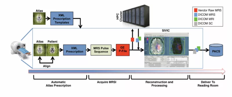
Our group created the workflow for obtaining 3D proton-1 (1H) MR spectroscopic imaging (multi-voxel MRS) datasets using an atlas-based automatic prescription, scanner reconstruction, and DICOM output at research scanners. Our recent research focuses on translating these tools and results to the broader neuro-oncology community and integrating them into the standard clinical workflow.
2. Develop Automatic, Rapid, High-resolution 1H MRSI using AI-based Approaches
We will automate the process of acquiring and generating accurate, high-resolution images of brain and tumor metabolism to aid clinicians in precisely identifying tumor boundaries using artificial intelligence (AI) approaches.
3. Develop novel deuterium (2H) MRS
2H MRS has been presented as a novel tool that can simultaneously detect glucose flux to lactate via anaerobic metabolism and glutamate/glutamine via mitochondrial metabolism following the administration of deuterated glucose (2H-glucose). We work on developing an imaging protocol to obtain reliable measures for evaluating patients with glioma (Warburg effect) and major depressive disorder (glutamate metabolism).
Opportunities: Volunteer Metabolic Imaging