Gliomas are the most common malignant primary brain tumors in adults. Our group is interested in developing, implementing, and applying a multi-modal MR protocol, including hyperpolarized carbon-13 ([1-13C]pyruvate, [2-13C]pyruvate), a new molecular imaging modality to provide real-time brain metabolism, and 1H MRSI to improve patient management. The research projects that our lab focuses on imaging glioma are shown below.
1. Characterize Spatial Heterogeneity in Astrocytoma
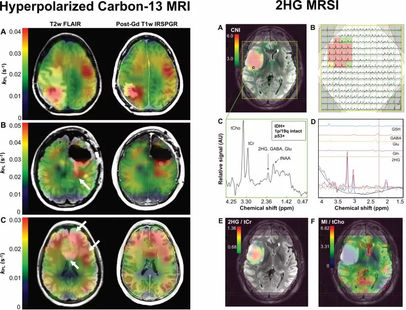
Although MRI is commonly used to monitor patients with glioma, the structural MR images commonly obtained for clinical diagnosis do not clearly define these tumors' boundaries. Our group works on characterizing spatial heterogeneity using dynamic hyperpolarized carbon-13 ([1-13C]pyruvate, [2-13C]pyruvate) and steady-state 1H 2-hydroxyglutarate specific hydroxyglutarate to improve the management of patients with astrocytoma.
2. Evaluate Cognitive Functioning in Astrocytoma
Although survival outcomes for lower-grade gliomas improve as diagnosis evolves, these patients eventually undergo resistance to treatment and often experience impaired cognitive function. Our group works on studying impairment in cognition and quality of life due to treatment and tumor progression using multi-modal neuroimaging.
3. Assess Early Response to Therapy and Tumor Burden in Astrocytoma
Early assessment of whether the tumor is responding or exhibiting signs of progression is crucial for optimizing clinical management and critical in defining endpoints in clinical trials. Our group is interested in enhancing the evaluation of novel treatments and assisting in patient management by using novel dynamic noninvasive hyperpolarized carbon-13 probes.